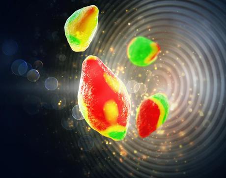
X-ray microscope technique lets scientists see inside lithium batteries

Researchers have developed an X-ray microscopy technique that will help scientists see what’s happening inside lithium-ion battery particles as they charge and discharge. Being able to see what’s going on will help scientists better understand how the batteries work, and how to make them better.
As reported in the journal Science, the scientists used the approach to image micron-sized battery particles as lithium ions migrate in and out of the particles. The images chronicle the evolution of the particles’ chemical composition and reaction rates at a nanoscale spatial resolution and a minute-by-minute time resolution.
Among the findings, the scientists discovered the charging process doesn’t play out uniformly on the surface of a particle, a phenomenon that likely curbs battery performance over time. This and other insights obtained from the imaging technique could help researchers improve batteries for electric vehicles as well as smartphones, laptops and other devices.
“The platform we developed allows us to image battery dynamics at the mesoscale, which is between a few nanometres and a few hundreds of nanometres,” said Will Chueh, a faculty scientist at the Department of Energy’s SLAC National Accelerator Laboratory, and an assistant professor of materials science and engineering at Stanford University, who led the research.
“This is a very difficult length scale to image in a functioning battery, but it’s critically important, because this is the scale that controls the fundamental processes involved in battery degradation and recharge time,” said Chueh.
The scientists developed the platform at the Advanced Light Source, which produces light in the X-ray region of the electromagnetic spectrum. The technique was implemented at two beamlines that offer high-performance scanning transmission X-ray microscopy (STXM), in which an extremely bright X-ray beam is focused onto a small spot.
Their specially designed ‘liquid electrochemical STXM nanoimaging platform’ uses the facility’s soft X-rays to image lithium iron phosphate particles as they charge (delithiate) and discharge (lithiate) in a liquid electrolyte. Their experimental set-up can image about 30 particles at a time.
In a real battery, thousands of these particles form an electrode, and positively charged lithium ions embed in the electrode as the battery charges. Ideally, the ions are inserted uniformly across the electrode’s surface. But this rarely happens, especially as a battery ages, which negatively affects performance.
“We now have a way to study this process in real time at the scale it’s occurring, which will help scientists better understand the process and possibly optimise it,” said David Shapiro, a physicist with the Advanced Light Source, who helped Chueh develop the technique at the facility.
“Our STXM-based platform provides the ability to image these electrochemical changes within a single battery particle,” Shapiro added. “And it offers the real-time resolution required to map the changes in the particles’ chemical composition and current densities, at a subparticle scale, as the particles lithiate and delithiate.”
Molecular Velcro coating boosts solar cell efficiency
Researchers have developed a new coating layer that enhances the durability and efficiency of...
Predictive AI model enhances solid-state battery design
ECU researchers are working on ways to make solid-state batteries more reliable with the help of...
Boosting performance of aqueous zinc–iodine batteries
Engineers from the University of Adelaide have enhanced aqueous zinc–iodine batteries using...


